Abstract
Recombinant, replication-competent rabies virus (RV) vaccine strain-based vectors expressing HIV type I (HIV-1) envelope glycoprotein (gp160) were developed from both a laboratory-adapted ( CXCR4-tropic) as from a primary (double tropic) isolate of HIV-1. An additional transcription stop/start unit within the RV genome was used to express HIV-1 gp160 in addition to the other RV proteins. HIV-1 gp160 protein was stably and functionally expressed, as indicated by fusion of human T-cell lines after infection with recombinant Rabies virus (RVs).
Inoculation of mice with the recombinant RVs expressing HIV-1 gp160 induced a strong humoral response directed against the HIV-1 coat protein after a single boost with recombinant HIV-1 gp120 protein. Furthermore, high neutralization titers of up to 1:800 against HIV-1 could be detected in mouse sera. These data indicate that a live recombinant RV, a rhabdovirus, expressing HIV-1 gp160 can serve as an effective vector for a vaccine against HIV-1.
Materials and methods
- Plasmid construction.
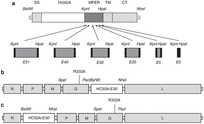
Two unique sites were introduced into the previously described RV cDNA page L16(17) upstream of the G (SmaI) and Ψ (NheI) gene by site-directed mutagenesis (GeneEditor, Promega) using primers RP11 5′-CCTCAAAAGACCCCGGGAAAGATGGTTCCTCAG-3 ′ and RP12 5′-GACTGTAAGGACYGGCTAGCCTTTCAACGATCCAAG-3′, resulting in plasmid PSN. BSN was the target used to introduce a new transcription Stop/Start sequence as well as a single BsiWI site using a PCR strategy.
First, two fragments were amplified by PCR from PSN using Vent polymerase (New England Biolabs) and the forward primers RP1 5′-TTTTGCTAGCTTATAAAGTGCTGGGTCATCTTAAGC-3′ or RP10 5′-CACTACAAGTCAGTCGAGACTTGGAATGAGATC-3′. The reverse primers were RP18 5′-TCTCGAGTGTTCTCTCTCCAACAA-3′ and RP17 5′-AAGCTAGCAAAACGTACGGGAGGGGTGTTAGTTTTTTTCATGGACTTGGATCGTTGAAAGGACG-3′.
RP17 contains an RV transcription stop/start sequence (underlined) and a BsiWI and NheI site (in italics). The PCR products were digested with NheI and ligated, and the 3.5 kb band eluted from an agarose gel. After elution from the gel, the band was digested with ClaI/MluI and ligated to PSN previously digested with ClaI/MluI. The plasmid was named ESPN.
HIV-1 gp160 genes, encoding the envelope protein of HIV-1 strains 89.6 and NL4-3, were amplified by PCR using Vent polymerase, forward primer 5′-GGGCTGCAGCTCGAGCGTACGAAAATGAGAGTGAAGGAGATCAGG-3′ containing sites PstI/XhoI/BsiWI (in italics), and the reverse primer 5′-CCTCTAGATTATAGCAAACCCCTTTCCAAG-3′ containing an XbaI site (in italics). The PCR products were digested with PstI and XbaI and cloned into pBluescript II SK(+) (Stratagene).
After sequence confirmation, the HIV-1 gp160 genes were excised with BsiWI and XbaI and ligated into ISBN, which had been digested with BsiWI and NheI. The resulting plasmids were named ESPN-89.6 and ISBN-NL4-3.
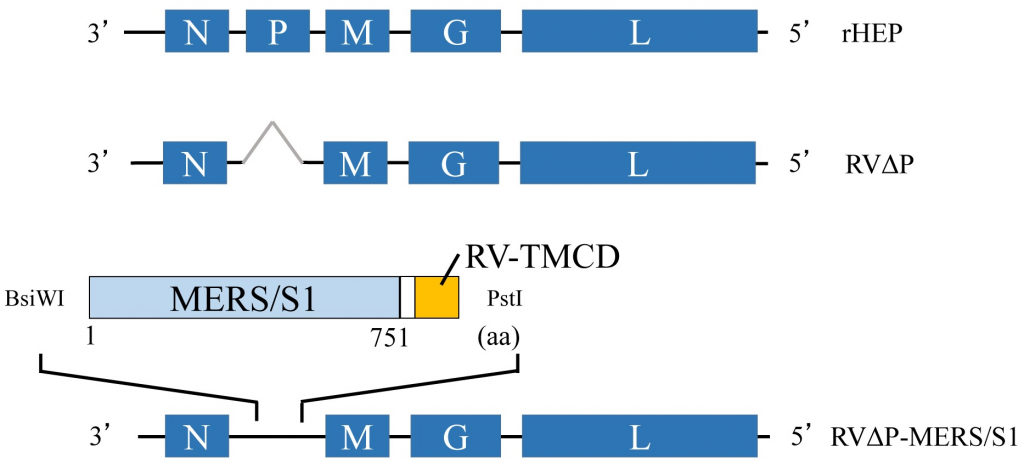
- Recovery of infectious RV from cDNA.
For rescue experiments of recombinant RVs, we used the vaccinia-free RV recovery system described previously. Briefly, BSR-T7 cells, stably expressing T7 RNA polymerase (a generous gift from S. Finke and K.-K. Conzelmann, Genzentrum, Munich) were transfected with 5 μg of cDNA from full-length RV plus plasmids encoding RV N, P, and L proteins (2.5, 1.25, and 1.25 μg, respectively), using a Ca2PO4 transfection kit (Stratagene) as directed by the supplier. Three days after transfection, tissue culture supernatants were transferred to fresh BSR cells and three days later infectious RV was detected by immunostaining against RV N protein (Centocor).
- One-step growth curve.
BSR cells (a clone BHK-21) were seeded in 60 mm dishes and infected 16 hours later (7 × 10⁶ cells) at a multiplicity of infection (moi) of 10 with SBN, SBN-89.6 or SBN-NL4-. 3 in a total volume of 2 ml. After incubation at 37°C for 1 hour, the inocula were removed and the cells were washed four times with PBS to remove any unabsorbed virus. Three millilitres of complete medium was added back and 100 µl of tissue culture supernatants were removed at 4, 16, 24 and 48 hours post-infection. Virus aliquots were titrated in duplicate on BSR cells.
- Immunization.
Groups of five 4- to 6-week-old female BALB/c mice obtained from The Jackson Laboratory were inoculated subcutaneously in both hind footpads with 106 foci-forming units of SBN, SBN-89.6, or 105 NL4-3 in DMEM. + 10% SFB. Three of five mice in each group were boosted intraperitoneally 3 months after infection with 10 μg recombinant gp41 (IIIB, Intracel, Issaquah, WA) and 10 μg recombinant gp120 (IIIB, Intracel) in 100 μL Freund’s complete adjuvant.
- Enzyme-linked immunosorbent assay (ELISA).
Recombinant HIV-1 gp120 (strain IIIB, Intracel) was resuspended in coating buffer (50 mM Na2CO3, pH 9.6) at a concentration of 200 ng/mL and plated onto 96-well MaxiSorp ELISA plates (Nunc) at 100 µl in each. good. After overnight incubation at 4°C, plates were washed three times (PBS, pH 7.4/0.1% Tween-20), blocked with blocking buffer (PBS, pH 7.4/ 5% milk powder) for 30 minutes at room temperature. and incubated with serial dilutions of sera for 1 hour.
Plates were washed three times, followed by the addition of horseradish peroxidase (H+L)-conjugated goat anti-mouse IgG secondary antibody (1:5,000, Jackson ImmunoResearch). After a 30 minute incubation at 37°C, the plates were washed three times and 200 µl of OPD (o-phenylenediamine dihydrochloride, Sigma) substrate was added to each well. The reaction was stopped by the addition of 50 µl 3M H2SO4 per well. The optical density was determined at 490 nm.

