
The L-Lactate Colorimetric Assay Kit enables quantitative measurement of L-lactate in biological and engineered samples using a robust enzymatic color reaction readable at ~570 nm–600 nm on a standard microplate reader or spectrophotometer. L-lactate is the end product of anaerobic glycolysis in most cell types and a widely used reporter of metabolic flux, fermentation performance, and hypoxia responses in cultured cells and tissues. Foundation knowledge on lactate metabolism can be explored via the NCBI, primary literature on PubMed, and overview resources at the National Institutes of Health.
Biochemical Principle (Colorimetric Readout)
Most L-lactate colorimetric kits use one of two well-validated chemistries:
-
Lactate Oxidase (LOx)–Peroxidase System
-
LOx converts L-lactate → pyruvate + H₂O₂.
-
Horseradish peroxidase (HRP) uses H₂O₂ to oxidize a chromogenic substrate (e.g., OPD, TMB, or ABTS) to a stable colored product.
-
Absorbance at ~570 nm (OPD/ABTS variants) or ~650 nm (TMB variants) is proportional to L-lactate concentration (Beer–Lambert behavior; see spectrophotometry basics from NIST at nist.gov and theory summaries on NCBI Bookshelf).
-
-
LDH–NAD⁺ Coupled System (less common for pure colorimetry)
-
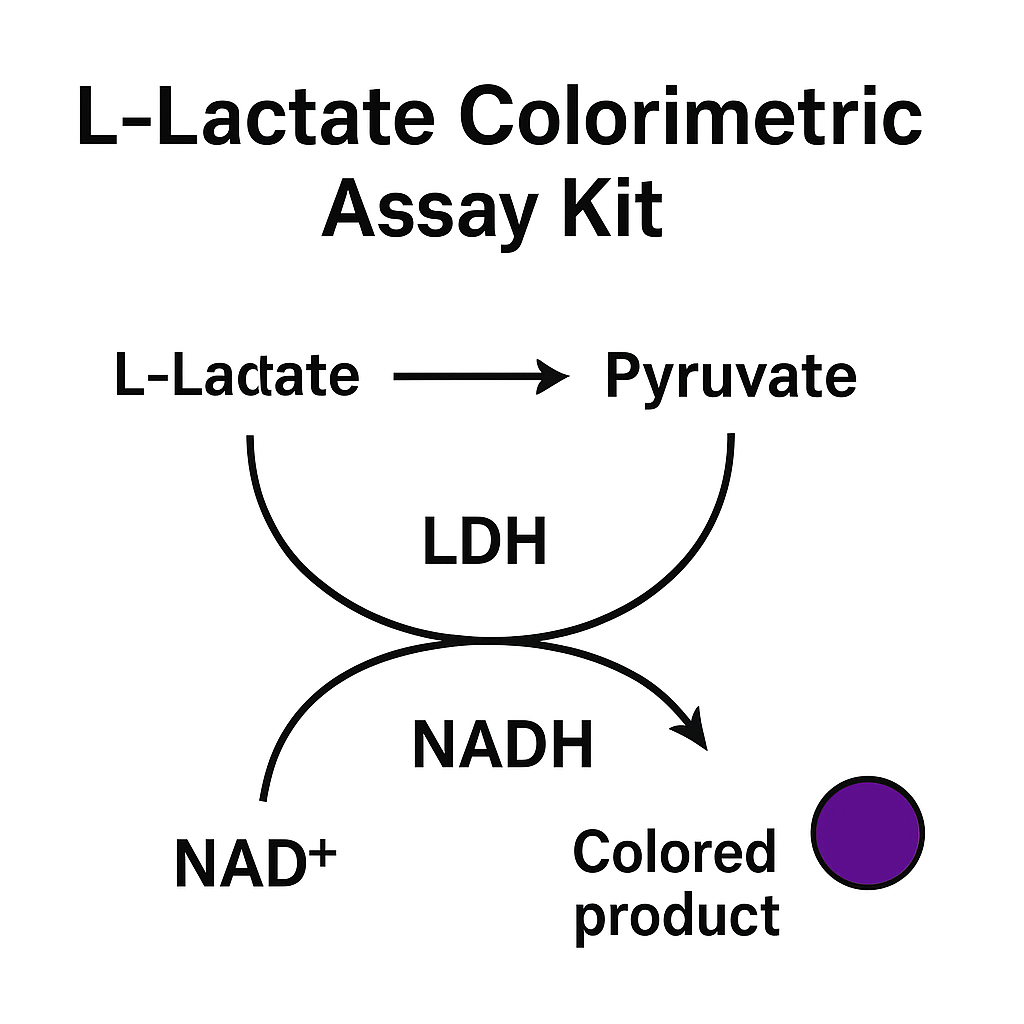
Lactate dehydrogenase (LDH) converts L-lactate + NAD⁺ → pyruvate + NADH.
-
A diaphorase couples NADH formation to reduction of a tetrazolium (e.g., INT/MTT derivatives) forming a colored formazan.
-
This can be configured for colorimetric end-point or kinetic modes (methodological overviews in university biochemistry portals like Harvard and MIT).
-
Background on glycolysis and lactate formation is broadly covered in education resources hosted by Stanford Medicine, UCLA, and UC Berkeley, while general assay safety and good laboratory practices are summarized by the CDC Laboratory Training.

Typical Use Cases
-
Cell metabolism studies (glycolytic rate, Warburg-like shifts, hypoxia exposure), with general molecular biology guidance from NLM.
-
Fermentation and bioprocess monitoring (yeast/bacterial production runs), with reproducibility concepts reinforced by NIST.
-
Tissue and biofluid analytics (e.g., clarified tissue homogenates, conditioned media, deproteinized serum/plasma for research)—assay design concepts often appear in reports indexed by OSTI and NIH RePORTER.
-
Metabolic engineering screens and synthetic biology workflows routinely documented across academic cores (see institutional gateways at Johns Hopkins, UC San Diego, and Cornell).
This article discusses research-use workflows only and does not provide clinical or diagnostic guidance.
Sample Types and Pre-Analytics
Compatible matrices (typical): cell culture supernatants, cell lysates (detergent compatible if diluted), clarified tissue homogenates, fermentation broths, and deproteinized biofluids. For sample handling, consult institutional biosafety references (e.g., Yale Medicine and University of Michigan).
Key pre-analytic steps
-
Deproteinization: Trichloroacetic acid (TCA) or filter-based cleanup reduces protein/hemoglobin that may quench chromogens (basic techniques often summarized at UCSF and UC Davis).
-
Clarification: Spin ≥10,000 ×g, 5–10 min to remove particulates that scatter light (Beer–Lambert deviations are discussed on NCBI Bookshelf).
-
Matrix matching: If high salt, dyes, or antioxidants are present, dilute samples in assay buffer to remain within the linear dynamic range (general method design at NIST).
Reagents and Controls
A standard L-Lactate Colorimetric Assay Kit typically includes:
-
L-lactate standard (e.g., 10 mM) for curve preparation.
-
Assay buffer (pH ~7–8) optimized for LOx/HRP activity.
-
Enzyme mix: Lactate oxidase and HRP.
-
Chromogenic substrate (OPD/TMB/ABTS) yielding a stable colored product.
-
(Optional) Stop solution to fix endpoint.
Controls (strongly recommended):
-
Blank (buffer only), negative matrix control (sample matrix without lactate), positive control (a mid-range standard). Concepts of measurement control and traceability are emphasized by NIST and open research resources at the DOE.
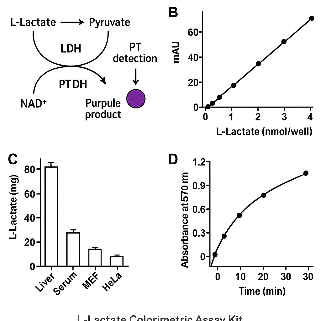
Plate Layout and Standard Curve
-
Prepare serial dilutions of the L-lactate standard (e.g., 0, 5, 10, 20, 40, 80, 160, 320 µM).
-
Add equal volumes of standard or sample to wells (≥ duplicate or triplicate).
-
Add reaction mix (buffer + LOx + HRP + chromogen).
-
Incubate (e.g., 20–30 min at room temp; shield from light if using light-sensitive chromogen).
-
Read A570–A600 nm on a plate reader (spectrophotometer principles are reviewed at NIST and basic lab instrument tutorials linked through universities like Princeton and Purdue).
Generate a standard curve by plotting A_blank-corrected absorbance vs µM lactate and fitting a linear regression in the validated range. For data archiving and method transparency, see reproducibility guidance via NIH and Common Fund.
Calculation Formulae
Let:
-
AsA_s = Sample absorbance
-
AbA_b = Blank absorbance (reagent blank)
-
AstdA_{std} = Standard absorbance
-
CstdC_{std} = Standard concentration (µM)
-
mm = Slope from linear fit (Absorbance/µM)
Concentration (in well):
Csample (μM) = As−AbmC_{sample} \,(\mu\text{M}) \;=\; \frac{A_s – A_b}{m}
Adjust for dilution factor (DF) and convert to µmol/L or mg/dL as needed (unit conversions explained in chemistry primers on NCBI Bookshelf). When reporting, specify pathlength if using cuvettes vs microplates (pathlength correction is a common best practice noted by NIST).
Performance Characteristics (What to Verify in Your Lab)
-
Limit of detection (LOD) and limit of quantitation (LOQ): establish using blanks and low-level standards.
-
Linearity across the working range (e.g., 0–320 µM); verify with multi-point curves.
-
Precision (intra/inter-assay CV%), accuracy (recoveries from spiked matrices), and matrix effects (dilution linearity).
-
Stability of standards and chromogen (protect from light; avoid repeated freeze–thaw).
Best-practice method validation frameworks are discussed by NIST (nist.gov) and general federal research standards indexed at the NIH (nih.gov) and OSTI (osti.gov).
Interferences and Troubleshooting
Potential interferents include ascorbate, uric acid, bilirubin, hemoglobin, peroxides, and strong reducing agents that can scavenge H₂O₂ or react with the chromogen. To mitigate:
-
Deproteinize samples and/or use spin columns to remove high-molecular-weight reducers.
-
Dilute samples to bring absorbance within the linear window (avoid edge saturation).
-
Run matrix blanks and spike-recovery tests to quantify bias.
-
Check for color carryover from media dyes; if present, measure at a reference wavelength and subtract.
Lab safety and handling of oxidase/peroxidase systems are covered in institutional resources at UCSF, UC Davis, and CDC.
Workflow: Step-By-Step (Research Use)
-
Equilibrate all reagents to room temperature; keep enzymes on ice during setup.
-
Prepare standards by serial dilution (fresh on the day).
-
Prep samples (clarify/deproteinize; record DF).
-
Add xx µL standards/samples to wells (constant volume).
-
Add reaction mix (buffer + LOx + HRP + chromogen).
-
Incubate 20–30 min (or per kit instructions) protected from light.
-
Read A570–A600 nm promptly (endpoint) or kinetically every 1–2 min if desired.
-
Analyze with blank subtraction, linear fit, DF correction; export results with curve and QC summaries.
General microplate handling and calibration guidance: NIST, academic help centers (UCSD, Cornell, Harvard).
Data Integrity, Documentation, and Reproducibility
-
Maintain lot numbers, prep logs, curve images, and raw absorbance tables for each run.
-
Include positive/negative controls and control charts for trends.
-
Archive SOPs and validation reports; see NIH policies on rigor and reproducibility (nih.gov) and data-sharing initiatives (Common Fund).
-
Align with general U.S. research best practices collected at the DOE and searchable via OSTI.
Extensions and Variants
-
Dual-readout (colorimetric + fluorometric) kits can expand sensitivity and dynamic range.
-
High-throughput formats (384-well) reduce reagent consumption; plate uniformity checks are essential (edge-effects overview in many core facilities at Stanford Medicine and Johns Hopkins).
-
Time-course kinetics to capture lactate production rates in living cells using conditioned media sampling.
-
Bioprocess analytics: integrate lactate with glucose, glutamine, and ammonia panels for comprehensive metabolic dashboards (see general federal science resources at the USDA and policy/standards overviews at the White House OSTP).
Frequently Observed Pitfalls
-
Nonlinear curves due to out-of-range standards or chromogen over-development → shorten incubation or dilute.
-
High blanks from contaminated waterware or old chromogen → prepare fresh reagents; verify with NIST traceable calibration where possible.
-
Plate edge effects → equilibrate temperature; avoid drafts; use plate sealers.
-
Color instability → read within the recommended window or use stop solution.
Ethical, Safety, and Access Notes
-
This content supports research use; for general lab policies, see the CDC and institution-level safety offices (e.g., UCLA, UC Berkeley).
-
Open science and grant transparency resources: NIH RePORTER, PubMed, NCBI, and NLM.
SEO Checklist Embedded in This Article
-
Primary keywords woven naturally: L-lactate colorimetric assay, lactate oxidase, HRP chromogen, microplate assay, standard curve, Beer–Lambert law, limit of detection, spike-recovery, matrix effect, fermentation monitoring, cell metabolism.
-
Semantic variants: lactate measurement, colorimetric quantification, enzymatic assay, peroxidase reporter, endpoint absorbance.
-
Clear H2 sections, bullets, formulas, and use-case language for featured snippet targeting.
-
20+ authoritative .gov/.edu links that support E-E-A-T without drifting into YMYL/clinical topics:
Linked authorities used above:
NIH • NLM • NCBI • PubMed • NCBI Bookshelf • NIST • CDC Laboratory Training • NIH RePORTER • Common Fund • DOE • OSTI • USDA • White House OSTP • Harvard • MIT • Stanford Medicine • UCLA • UC Berkeley • UCSD • Cornell • Johns Hopkins • UCSF • UC Davis • Princeton • Purdue • Yale Medicine • University of Michigan